


Robotic TME with Coloanal Anastomosis. Technical Aspects
April 22, 2012
Paolo Pietro Bianchi (Grosseto – Italy) Wanda Petz (Milano – Italy) L. Casali, M. Parodi, D. Belotti
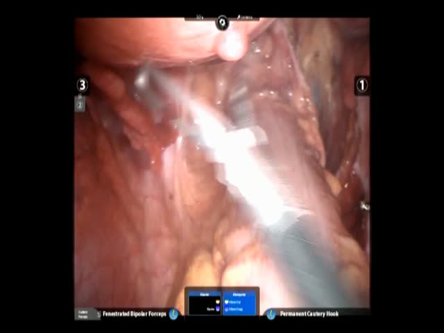
SUMMARY: The video shows a robotic rectal resection with manual colo-anal anastomosis for adenocarcinoma of the lower rectum. A 47 years old woman was diagnosed with a cT2N0 adenocarcinoma of the lower rectum located 1 cm from the dentate line. Preoperative assessment was performed with colonoscopy with biopsy, thoraco-abdominal CT scan, pelvic MRI and echoendoscopy. The patient was positioned in dorsal decubitus with open legs and the operating table was in Trendellemburg position and rotated to the right. Six trocars were inserted: one right paraumbilical for the optical system, three robotic trocars in right iliac fossa, left iliac fossa and epigastrium and two accessory trocars in the right flank. After laparoscopic exploration of the abdominal cavity with hepatic ultrasonography, the robotic arms were connected, the cart coming from the patient’s left flank. First operative step was splenic flexure mobilization, performed with a medial-to-lateral approach with isolation and section of the inferior mesenteric vein, opening of the lesser sac by sectioning the root of transverse mesocolon, and colo-epiploic detachment from the right to the left. Inferior mesenteric artery was then isolated and sectioned at its origin. Mesorectal excision started posteriorly to the arterial plane and was conducted until the muscular pelvic floor. The perineal phase of the surgery consisted in a mucosectomy of the lower rectum starting 2 cm distally from the tumour; the inter-sphinteric plane was then reached and the dissection proceeded cranially until reaching the abdominal level. The rectum was completely dissected and extracted through the anus; proximal resection was performed on the descending colon; after realizing a colic J pouch, a manual end-to-end anastomosis was performed between the pouch and the external sphincter. Operating time was 5 hours; postoperative course was uneventful, first bowel movements were observed on second postoperative day (POD) through the lateral ileostomy and the patient was discharged on 7th POD. Final histopathological examination confirmed a T2N0 adenocarcinoma with 32 harvested lymph nodes. Robotic surgical approach to the lower pelvis may increase the precision of rectal dissection and overcome technical difficulties of conventional laparoscopy.