


Robotic low anterior resection with fluorescence imaging
August 31, 2012
M.Hellan
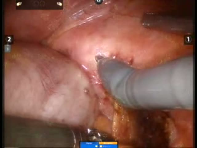
This is a male patient with a BMI of 34 and a large anterior midrectal cancer. Due to his obesity a laparoscopic medial to lateral approach is used for mobilization of the left sided colon. A high ligation of the IMA and IMV is performed. The Da Vinci Si system with three working arms is utilized for a total mesorectal excision. First the mesorectum is mobilized posteriorly and laterally all the way to the pelvic floor. The tumor appears attached to the seminal vesicles. Therefore the vesicles are resected en-bloc with the rectum. Douvillier?s fascia is divided distally and the low rectum is divided with 2 purple staple loads. The mesentery is divided all the way to the edge of the distal sigmoid colon in preparation for transection. 10 mg of indocyanin green (ICG) dye is injected intravenously and the vision mode is switched to fluorescence imaging. The perfusion of the rectal stump as well as the proximal sigmoid is assessed and deemed adequate under fluorescence imaging. The colon is now divided at the planned transection line through a small supra-pubic incision. The rectal anastomosis is completed with an EEA 29 stapler.