Robotic Resection of Paracaval Leiomiosarcoma
April 26, 2012
Paolo Pietro Bianchi (Grosseto – Italy) Wanda Petz (Milano – Italy) L. Casali, M. Parodi, D. Belotti
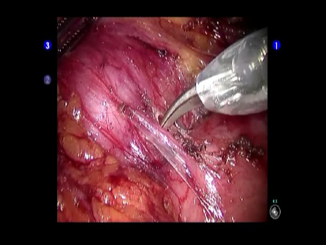
The robotic approach can help to perform fine dissection of lesions even if located in difficult anatomical regions. The video shows a robotic resection of a paracaval mass of unknown origin. The patient was a 50 year-old male with silent medical history who presented with a 7 cm paracaval mass of unknown origin. With the patient in left lateral position, five trocars were inserted: one optical right paraumbilical trocar, three 8 mm robotic trocars in the left hypocondrium and one in the right flank and 2 accessory laparoscopic trocars, paraumbilical and epigastric. After intraoperative ultrasonography, the posterior parietal peritoneum was opened inferiorly to the liver; the lesion was dissected from the duodenum and carefully from the vena cava and from the right renal vein. The specimen was then removed in a plastic bag enlarging the optical access. Operating time was 180 minutes with negligible blood loss. No intraoperative complications occurred. Oral feeding was reintroduced in first post operative day (POD) and the patient was discharged in second POD without complications. Histopathological examination revealed a low grade leiomyosarcoma. The robotic assistance can help to realize fine surgical dissection and to perform conservative surgery also in difficult retroperitoneal tumors.


