Robotic Radical Thymectomy
May 5, 2012
Pier C. Giulianotti (Chicago – USA) Francesco Bianco ( Chicago – USA) K. Venkata
Disease: Myasthenia gravis.
Age: 37
ASA score: 1
Histology: Involuted thymic tissue with focal calcifications
History: A 37-year-old Caucasian female who has myasthenia gravis with complaints of arm and leg weakness, fatigue and some difficulty chewing.
Description: Trocars: introduction of a 5-mm trocar in the fifth intercostal space, very lateral, close to the posterior axillary line. The right lung is excluded by the anesthesiologist when a low pressure is reached. Two more trocars, one in the third intercostal space anterior axillary line and the other is in the fifth intercostal space, always in the anterior axillary line, are placed. The posterior Trocar is upsized to 10 for the scope and another 5-mm port is placed between the camera port and the upper port in the third intercostal space.
Steps
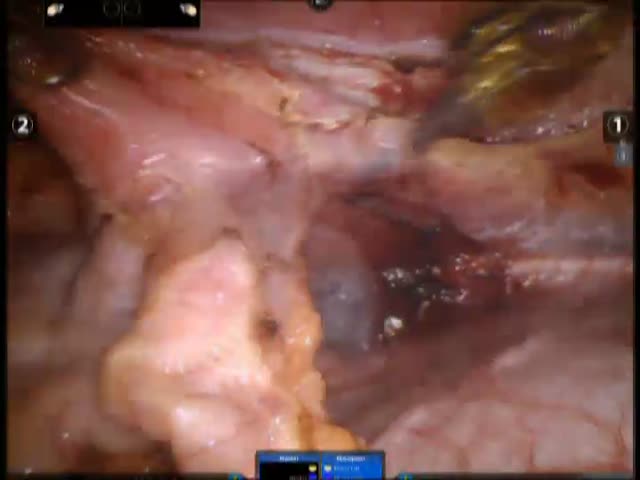
1 – Opening of the Pleura in the anterior mediastinum. The right phrenic nerve and the internal mammary vessels are identified and preserved.
2 – The thymus is completely isolated and dissected without breaking the capsule, removing some pericardial fat as well and the horns of the thymus that
are particularly long, mainly on the right side. The two thymic veins that are going to the innominate vein are identified and those veins are clipped with the Hemo-loch clips and then transected. The left pleura is open and the phrenic nerve on the other side identified. A radical thymectomy is completed. Blood loss is less than 50 mL.


