


Robot-assisted Right Hepatectomy and Cholecystectomy
May 12, 2012
Pier C. Giulianotti (Chicago – USA) Subhashini Ayloo (Newark – USA)
Disease: Giant liver hemangioma, segment 7/6
Age: 46
Previous Surgeries: Hysterectomy, tonsillectomy and aneurysm clipping for cerebral aneurysm 8 years before.
Histology: Cavernous hemangioma (10 x 5 x 5 cm), subcapsular, with foci of thrombosis, organization and sclerosis
History: Patient developed chest pain approximately 3 to 4 months before with spasms radiating to her back. She had further imaging performed and was found to have a 9 cm hemangioma of her liver. An MRI demonstrated a 10.2 cm lesion in the superior aspect of the right hepatic lobe extending toward the inferior aspect.
Description: Trocars: A 5-mm cannula is inserted in the left upper quadrant. One 10/12 port is placed in the right pararectal line on the right side of the umbilicus, then
another 10/12 on the left side of the umbilicus. Three 8-mm ports are placed
one in the right upper quadrant, two in the left upper quadrant.
Steps
1 – Ultrasound evaluation of the liver using a laparoscopic probe and confirming the lesion located in the segment 7 and segment 6, and almost reaching the right subhepatic vein.
2 – Cholecystectomy: the fluorescence is very useful in this case because we are able to identify a small aberrant duct that is from the gallbladder bed going to the cystic duct. This anomaly is clearly recognized and recorded. Finally, the small duct is transected having the confirmation of the nature and a stitch of Prolene 4-0 to secure the proximal stump of the duct, even though the small duct will be removed together with the specimen.
3 – Hilum dissection: Identification of the right hepatic artery. Whith the use of a bulldog clamp, we ensure that the discoloration connected to the ischemia is limited to the right side. The two branches of the right hepatic artery are doubly ligated and the portal vein underneath is exposed. The right trunk of the portal vein is completely isolated. The right portal trunk is encircled and then with the suture of 3-0 Prolene and one clip, the portal vein is transected.
The bifurcation of the bile duct is identified and confirmed with fluorescence.
The right lobe is lifted with the fourth arm and the vena cava is exposed. The main discharge is the right hepatic vein plus another big branch that is joining the right hepatic vein coming from the segment 6. As an extracapsular control looks suboptimal, we are deciding to control the hepatic veins
inside the capsule.
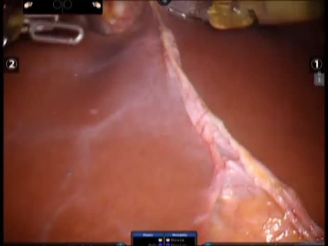
4 – Transection of the parenchyma using the Harmonic and perfecting hemostasis either with the bipolar or Prolene stitches. The central area is characterize multiple deep connections with the hepatic vein system requiring a stapler.
The overall blood loss during the hepatectomy has been containing no more than 350 cc of blood. The patient was fine for the entire procedure, stable, and now she is sent stable and in good condition to the medical ICU.